The result adds long-term evidence for supplemental ultrasound, but it does not create a worldwide screening rule. The study did not use breast cancer mortality as an endpoint, and its age range, Japanese setting and two-round design limit how far the result can be applied.
New breast changes need prompt assessment
Screening is intended for people without symptoms. The World Health Organization (WHO) lists a new breast lump or thickening, a change in breast size or shape, skin dimpling or redness, a nipple or areola change, and abnormal or bloody nipple fluid as possible breast cancer symptoms.
A person with one of these changes should seek clinical assessment promptly rather than wait for a screening invitation or the next routine mammogram. Most breast lumps are not cancer, so a symptom is not a diagnosis; it is a reason to move from routine screening to diagnostic assessment.
Mammography evidence does not produce one global schedule
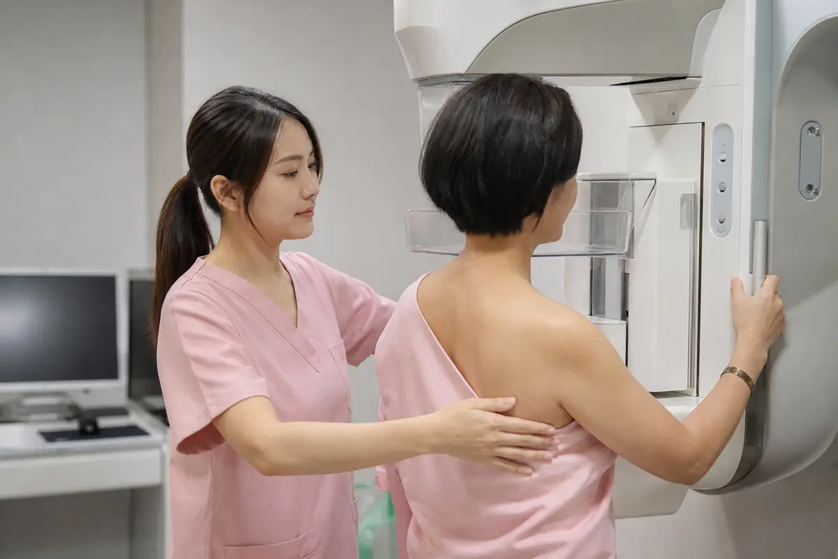
The International Agency for Research on Cancer (IARC) found sufficient evidence that mammography screening reduces breast cancer mortality among women ages 50 to 69. Its 2016 evaluation estimated an average 24 percent reduction among women invited to service screening and about 40 percent among those who attended. The gap shows how the population counted changes the result.
WHO describes screening as mammography in an apparently healthy population, usually women ages 50 to 69. The US Preventive Services Task Force instead recommends mammography every two years from ages 40 to 74 for people at average risk in the United States.
Neither range is a worldwide eligibility or coverage rule. The applicable starting age, interval and route to follow-up come from the program or clinical guidance used in the country where care is received.
Mammography also has harms. The US task force lists false-positive and false-negative results, extra imaging and biopsy, overdiagnosis and radiation exposure; IARC found that the radiation-related cancer risk was outweighed by the mortality benefit for women ages 50 to 74, but that conclusion does not erase the other costs of screening.
Density describes the mammogram, not a disease
Dense breasts are not an abnormal condition or a cancer diagnosis. Density can change with age and other factors, and a radiologist's classification is an assessment at the time of the mammogram.
A dense result does not make mammography pointless, and a negative mammogram does not guarantee that no cancer is present. The decision on added imaging depends on the complete report, the person's wider risk profile and the guidance in the health system providing care.
Ultrasound finds more cancers and more false positives
In 2024, the US task force found insufficient evidence to recommend for or against supplemental ultrasound or magnetic resonance imaging (MRI) after a negative mammogram in people with dense breasts. The European Commission Initiative on Breast Cancer conditionally advised organized screening programs against routine automated or hand-held supplemental ultrasound for asymptomatic women with dense breasts and a negative mammogram, citing low or very low certainty. These positions do not mean that supplemental imaging is never used; they mean that density alone has not produced a universal routine schedule.

Higher-risk and special populations need separate pathways
The average-risk schedules above do not cover every person. The US task force excludes people with a high-risk genetic marker or syndrome, high-dose chest radiation at a young age, previous breast cancer or a high-risk breast lesion from its average-risk recommendation.
The adult screening ranges cited here do not apply to children and do not establish one schedule during pregnancy or breastfeeding. People in those groups should use clinician-led assessment if a breast symptom appears rather than apply an adult routine screening interval.
NCI says postmenopausal hormone therapy is associated with higher breast density. The cited sources do not provide a general basis for changing a prescribed medicine solely because a mammogram reports dense tissue; medicine changes and screening decisions require clinical judgment.
A dense result changes the questions, not the diagnosis
A review of a mammogram report should separate the density category from the radiologist's assessment and recommended follow-up. Density alone is neither a cancer diagnosis nor an automatic order for ultrasound.
After a negative mammogram, the case for another imaging test depends on total breast cancer risk, the density category, prior findings and local guidance. An abnormal screening result or a new symptom follows a diagnostic pathway instead of the next routine screening interval.
The source article presented a 41 percent mortality reduction as a general screening effect. IARC's closest international evaluation produced different estimates for invitation and attendance within specified age groups, so APPI News has not treated 41 percent as a worldwide personal benefit. The source's stage-specific survival percentages were also omitted because no international dataset with matching stage definitions and population was identified.
Frequently asked questions
Does a dense-breast result mean cancer?
No. It describes the mix of tissue seen on a mammogram, although it can both mask cancers and raise breast cancer risk.
Can breast density be identified by touch?
No. NCI says a radiologist determines density from a mammogram, not from a self-exam or clinical breast exam.
Does dense tissue make mammography useless?
No. Mammography remains the core population screening test, but it is more likely to miss a cancer when tissue is dense.
Should everyone with dense breasts add ultrasound?
No universal rule supports routine ultrasound for everyone with a dense result and an otherwise negative mammogram. The potential for added detection has to be weighed against false positives, more imaging and biopsy.
What screening age and interval apply?
There is no single worldwide schedule. Eligibility and intervals should come from the program or clinical guidance used where the person receives care.
Should a new lump wait until the next mammogram?
No. A new lump, nipple change, abnormal nipple fluid or skin change needs clinical assessment rather than waiting for routine screening.
Sources and further reading
- Cumulative incidence of advanced breast cancer in women aged 40–49 years in J-START(The Lancet)
- Breast cancer(World Health Organization)
- Breast cancer screening: Evaluation(International Agency for Research on Cancer)
- Recommendation: Breast Cancer: Screening(US Preventive Services Task Force)
- Dense Breasts: Answers to Commonly Asked Questions(US National Cancer Institute)
- Mammography followed by ultrasonography compared with mammography alone(Cochrane)
- Tailored screening with automated breast ultrasound(European Commission Initiative on Breast Cancer)